Crouzon Syndrome
In This Article
Click to jump to your selected section.
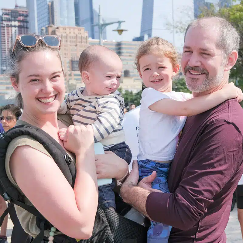
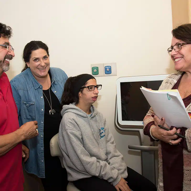
Meet a member of the myFace Community
Craniofacial Conditions > Crouzon Syndrome
Crouzon Syndrome
What is Crouzon Syndrome?
Crouzon syndrome is one of several types of craniosynostosis – a condition that results from the premature fusion of one or more of the seams (sutures) of the skull bones. This means that the cranial sutures, or the joints between the skull bones, have closed too early, resulting in skull and facial malformations.
The condition was first described in 1912 by French physician Octave Crouzon, who observed patients with characteristic skull malformations, facial anomalies, and proptosis (bulging of the eyes from their normal position).
Prevalence of Crouzon Syndrome
Often considered the mildest form of the various craniosynostosis syndromes, Crouzon syndrome is estimated to occur in 1 in 60,000 newborns and appears to affect males more often than females. Crouzon syndrome accounts for about 5% of all craniosynostosis patients, and is the second most common craniosynostosis syndrome (out of over 100 such identified syndromes), affecting about 5,000 people in the United States.
Crouzon Syndrome is often referred to as:
- Craniofacial dysostosis
- Craniostenosis (Crouzon type)
- Crouzon craniofacial dysostosis
Causes of Crouzon Syndrome
Crouzon syndrome is a rare genetic disorder that is caused by mutations in the Fibroblast Growth Factor Receptor (FGFR)-2 and -3 genes, which are located on chromosome 10. More than 90% of Crouzon syndrome cases are caused by various mutations in the FGFR-2 gene, in which 50 unique mutations have been identified to date. The remainder of cases are mostly caused by a specific mutation in FGFR-3, which is characteristically associated with acanthosis nigricans. Acanthosis nigricans symptoms include dark and thickened velvety skin which appears in body folds and creases, such as in the armpits and groin.
Crouzon Syndrome and Genetics
Both FGFR-2 and FGFR-3 are protein receptors that play a role in craniofacial bone formation: during embryonic development, these receptors are involved in the creation of osteoblasts, which are cells that are responsible for bone formation.
The mutated versions of these receptors are abnormally active and accelerate the differentiation of osteoblasts, which leads to the premature fusion of the sutures in the newborn’s skull.
For example, the fusion of the coronal and sagittal sutures causes the bones forming the forehead (frontal bones) to join prematurely to the sides of the skull (parietal bones).
Although Crouzon syndrome is associated with mutations in the FGFR-2 or -3 genes, it can present in different ways in different patients; while some patients may appear largely unaffected, others may have severe anomalies. An individual can acquire a mutation in these genes in one of two ways. In about 50% of cases, patients inherit Crouzon syndrome in an autosomal dominant pattern. This means that they have inherited at least one copy of the mutated gene from one of their parents.
In autosomal dominant conditions, children of an affected parent have a 50% chance of inheriting the condition. In the remaining cases, the mutation is the result of a “de novo” (“new”) mutation, meaning it appears spontaneously for the first time in the affected individual, without any prior family history. The causes of these de novo mutations are largely unknown, but some studies have suggested a link with advanced paternal age.
Signs & Symptoms of Crouzon Syndrome
Premature fusion of cranial sutures (or “joints” between the various bones of the skull) during the first year of life leads to restricted growth and development of the skull, face, brain, and central nervous system. Thus, the clinical features become increasingly apparent during the first 1-2 years of life and can progress until age three. However, there can still be variability in the presentation, as some patients may show prominent signs at birth, while others may not manifest any changes until early childhood. Some people with Crouzon syndrome may have very mild facial changes, while others are more severe.
Read more about the Signs & Symptoms of Crouzon Syndrome →
Eye Abnormalities and Crouzon Syndrome
Ocular (eye) abnormalities are also frequently seen in Crouzon patients. Due to the early fusion of the sutures in the skull, the orbits (or eye sockets) are adversely affected. This causes the characteristic proptosis, or the protrusion of the eyeballs, seen in Crouzon patients. This can lead to dry eyes or excessive tearing and tends to make patients more susceptible to inflammation of the surface of the eye (keratitis) as well as conjunctivitis or “pink eye.” The abnormal orbital bone formation can result in an altered arrangement of the muscles behind the eyes, leading to strabismus (“crossed” or misaligned eyes) as well as amblyopia (“lazy eye”).
Glaucoma and Crouzon
Crouzon patients are also at higher risk for conditions that affect vision such as refractive errors and glaucoma. Refractive errors occur when the eye is misshapen, which affects the ability of light from focusing appropriately onto the retina; examples of refractive errors are near- and far-sightedness as well as astigmatism. Another common vision problem is glaucoma, which occurs when there is an abnormality in the eye’s drainage system. This can lead to the buildup of fluid and increased pressure inside the eye, which can ultimately result in damage to the optic nerve.
Comparison to Apert Syndrome
Although it shares overlapping features with other craniosynostosis syndromes such as Apert syndrome or Pfeiffer syndrome, it is important to note that patients with Crouzon syndrome typically have normally formed hands and feet. Patients with Crouzon syndrome do not show syndactyly, which is the webbing of the fingers or toes, that is commonly seen in Apert syndrome. They also do not have short broad big toes or thumbs, which are characteristic of Pfeiffer syndrome.
Diagnosing Crouzon Syndrome
It is possible to detect Crouzon Syndrome in a prenatal ultrasound, but it is more commonly diagnosed at birth or within the first year of life when the classic “crouzonoid features” are observed.
Physical signs of Crouzon Syndrome include:
- Craniosynostosis
- Midface Hypoplasia: underdevelopment of the eye sockets, cheekbones, and upper jaw
- Proptosis, or “bulging eyes”
- Beaked Nose
In patients with a known family history of Crouzon syndrome, these features are generally enough to confirm a diagnosis. However, in those with a spontaneous mutation, genetic testing and other imaging tests may be necessary, especially as the physical features may not be clear initially in the newborn.
Additional radiology tests may be needed to confirm the diagnosis including x-ray, CT, or MRI scans to image the bone structures and brain. If the diagnosis is still not confirmed based on history, clinical features, and imaging, molecular testing can be performed to assess whether the FGFR-2 or –3 mutations associated with Crouzon syndrome are present, which would help to distinguish it from other craniosynostosis syndromes.
Crouzon Syndrome Treatments & Procedures
Like many other craniofacial conditions, the management and treatment of Crouzon Syndrome depends on the features and severity of the appearance-related and functional impact on each individual patient. Aside from a pediatrician, a multidisciplinary team of craniofacial specialists, including oral maxillofacial surgeons, plastic surgeons, neurosurgeons, otolaryngologists (ENT), and ophthalmologists will likely be required to manage the patient.
Surgical Interventions
Surgical interventions are the main form of treatment for most children to prevent the most serious consequences of abnormal cranial development, including:
- Increased intracranial pressure
- Blindness
- Intellectual disabilities
- Hearing loss
- Respiratory and breathing problems
Generally, experts agree that early intervention before the age of 1 year results in lower risk of cognitive disabilities, airway obstruction, and visual problems. Surgery can take place in a staged or a combined approach.
Read more about Crouzon Syndrome Treatments & Procedures →
Living with Crouzon Syndrome
With proper care, patients with Crouzon syndrome typically have normal life spans. However, some long-term functional issues and complications may still persist.
Functional Challenges
The three most common functional challenges related to Crouzon syndrome are visual impairment, breathing issues, and hearing loss, as follows:
Visual Impairment
Because of the orbital proptosis (or bulging of the eyes) in Crouzon syndrome, patients have difficulty protecting the eye, leading to an increased risk of damage, inflammation, or infection to the eye, which can result in loss of vision. Also, due to increased intracranial pressure, the optic nerve can develop edema or swelling, resulting in vision loss. Thus, ocular development needs to be monitored carefully in the long-term, and regular monitoring for increased intracranial pressure should be done by using ophthalmologic and radiology exams until at least age 8.
Breathing Problems
Due to the underdevelopment of the jaw structures, many patients can develop obstructive sleep apnea (OSA). Patients with obstructive sleep apnea can experience respiratory depression and decreased oxygenation during sleep.
Read more about Breathing Problems associated with Crouzon Syndrome →
Hearing Loss
Due to abnormal growth and shape of the skull in Crouzon syndrome, deafness can occur when neural signals from the inner ear cannot be transmitted to the brain properly. Also, patients with Crouzon syndrome and other similar syndromes are at increased risk for recurrent otitis media (or middle ear infections), which can result in hearing loss. Hearing loss can occur in up to 50% of patients with Crouzon syndrome. Thus, proper care with an audiologist and other specialists are needed to monitor hearing, especially for those with recurrent infections, and to ensure the patient has hearing aids and other assistive devices to support proper speech development, learning, and social and emotional development. Children may also need support from ongoing speech and language therapy.
Potential Complications of Crouzon Syndrome
Increased intracranial pressure
Increase in the pressure inside the skull is the most serious complication of Crouzon syndrome, especially in the absence of a surgical intervention – such as open cranial vault remodeling – early in life to prevent this. In fact, in cases of untreated Crouzon syndrome, the incidence of increased intracranial pressure episodes has been reported to be almost 63%, with potential consequences for vision loss, blindness, and impaired cognitive development.
In Crouzon syndrome, the causes of increased pressure in the skull include hydrocephalus (increased fluid in the brain), airway obstruction, and intracranial venous hypertension (increased blood pressure in the veins within the brain). On average, the first episode of increased intracranial pressure occurs around 1.4 years old, and about half of these patients go on to experience a second episode about 1.4 years after the initial episode. Once increased intracranial pressure is detected, urgent treatment should be initiated based on the cause.
For example, in the case of hydrocephalus, a ventriculoperitoneal shunt can be placed to drain the excess fluid. If airway obstruction is the cause of increased intracranial pressure, the child may require an airway inserted into the nose and pharynx (a nasopharyngeal airway), initiation of continuous positive airway pressure to provide additional oxygenation, or may need to undergo surgery to remove the adenoids and tonsils to help open the airways. Lastly, if the cause is intracranial venous hypertension, the patient should be treated with a posterior vault expansion surgery (see above).
Chronic neurologic conditions
Patients with Crouzon syndrome may occasionally experience ongoing neurologic symptoms. For example, 25% of patients report having chronic headaches, and about 10% have recurring seizures or epilepsy. Chronic headaches can be associated with elevated intracranial pressure – but not always. In rare cases, it may also be caused by a persistent leakage of cerebrospinal fluid, which may have been caused by accidental puncture during spinal anesthesia during prior surgeries. The cause of seizures in Crouzon syndrome is unclear, but it may be related to the structural brain abnormalities caused by skull formation and hydrocephalus (excess fluid around the brain). Patients with seizures will need to be managed by a neurologist, who will use medications to minimize the occurrence of seizures as much as possible. Patients may only require one medication or sometimes combinations of medications to achieve adequate control.
Mental and emotional health challenges
As in patients with other craniofacial anomalies, children with Crouzon syndrome may suffer from self-esteem and confidence issues related to their appearance and chronic medical conditions. Visual and hearing impairments may hinder their social interactions, and they may experience social isolation or even bullying. Thus, ongoing psychosocial support from social workers and psychologists are important to help children with Crouzon syndrome and their families maintain mental and emotional wellness throughout the time of diagnosis and ongoing treatment. Special services can also help children adjust socially and ensure effective learning at school.
Developmental delay
Although most patients with Crouzon syndrome have normal intellectual ability, about 15% experience some mild to moderate developmental delay in their language, motor, or cognitive skills. Thus, the multidisciplinary treatment team should include a neuropsychologist who can evaluate a child for any developmental or cognitive delays and recommend interventions that can facilitate development.
Meet a Member of the myFace Community
myFace Provides Support for People Living with Crouzon Syndrome and Other Craniofacial Conditions
myFace provides a variety of direct services and educational programs to help meet the needs of the craniofacial community, including:
myFace provides a variety of direct services and educational programs to help meet the needs of the craniofacial community, including:
(1) myFace Virtual Support Groups provide counsel and community for individuals and their families living with facial differences. These help participants to feel safe, supported, and never alone. myFace offers Support Groups for adults, adolescents, and both English-speaking and Spanish-speaking parents.
(2) Parent Guides provide resources and guidance to assist parents as they support their child’s journey.
(3) Transforming Lives Webinar Series offers webinars with craniofacial experts on topics of interest to the community.
(4) myFace, myStory Podcast Series offers monthly interviews and roundtable discussions on topics of interest to the craniofacial community.
(5) myFace’s Guide to Craniofacial Surgeries serves as a guide to patients’ surgical experience, providing in-depth information on some of the most common craniofacial surgeries.
(6) myFace | Your Impact Newsletter provides the community with regular updates.
(7) myFace Family Apartments provide free housing for out-of-town families seeking treatment in New York City.
(8) myFace Wonder Project School Assemblies provides students with the tools they need to unite against bullying and to implement acts of kindness in their schools and in their communities.
(9) myFace Events provide opportunities for the craniofacial community to gather together.
References
- Abu-Sittah GS, Jeelani O, Dunaway D, Hayward R. Raised intracranial pressure in Crouzon syndrome: incidence, causes, and management. J Neurosurg Pediatr. 2016; 17: 469-475. (Full online access: https://thejns.org/pediatrics/view/journals/j-neurosurg-pediatr/17/4/article-p469.xml?tab_body=pdf-25465)
- Al-Namnam NM, Hariri F, Thong MK, Rahman ZA. Crouzon syndrome. J Oral Biol Craniofac Res. 2019; 9(1): 37-39. (Full article online: https://www.ncbi.nlm.nih.gov/pmc/articles/PMC6128172/)
- Bhattacharjee K, Rehman O, Venkatraman, et al. Crouzon syndrome and the eye: an overview. Indian J Ophthalmol. 2022; 70(7): 2346-2354.
- Conrady CD, Patel BC. Crouzon syndrome. StatPearls. Updated August 8, 2022. https://www.ncbi.nlm.nih.gov/books/NBK518998. Accessed November 6, 2022.
- Craniosynostosis syndromes. UpToDate. Reviewed October 2022. https://www.uptodate.com/contents/craniosynostosis-syndromes. Accessed November 6, 2022.
- Crouzon syndrome. Boston Children’s Hospital. https://www.childrenshospital.org/conditions/crouzon-syndrome. Accessed November 6, 2022.
- Crouzon syndrome. NORD. https://rarediseases.org/rare-diseases/crouzon-syndrome/ Accessed November 6, 2022.
- Crouzon syndrome. NIH Genetic and Rare Diseases Information Center. https://rarediseases.info.nih.gov/diseases/6206/crouzon-syndrome. Accessed November 6, 2022.
- De Jong T, Toll MS, de Gier HHW, et al. Audiologic profile of children and young adults with syndromic and complex craniosynostosis. Arch Otolaryngol Head Neck Surg. 2011; 137(8): 775-778. (Full article online: https://jamanetwork.com/journals/jamaotolaryngology/fullarticle/1106416)
- Ko JM. Genetic syndromes associated with craniosynostosis. J Korean Neurosurg Soc. 2016; 59(3): 187-191. (Full article online: https://www.ncbi.nlm.nih.gov/pmc/articles/PMC4877538)
- McCarthy JG, Warren SM, Bernstein J, Burnett W, Cunningham ML, Edmond JC, et al. Parameters of care for craniosynostosis. Cleft Palate Craniofac J. 2012;49:1S–24S
- Surgery for syndromes: surgical procedures for craniofacial syndromes. https://www.seattlechildrens.org/clinics/craniofacial/services/surgery-for-syndromes/ Accessed December 3, 2022.
In This Article
Click to jump to your selected section.
Meet a member of the myFace Community