Apert Syndrome
Craniofacial Conditions > Apert Syndrome
What is Apert Syndrome?
The gene responsible for Apert Syndrome is found on chromosome 10, known as Fibroblast Growth Factor Receptor 2 (FGFR2) (Charkins, 1996). Changes or mutations in this FGFR2 gene disrupt normal signaling in the body, causing the premature fusion of cranial sutures and abnormal bone development, which are hallmark features of Apert syndrome. By understanding the genetic roots of the condition, we not only improve our ability to diagnose it accurately but also open doors for targeted treatments to ease its impact.
Apert Syndrome
Overview: Also called acrocephalosyndactyly, Apert Syndrome is a genetic condition where the bones of the skull, hands, and feet fuse together.
It’s marked by abnormalities in the shape of the skull, face, teeth, and limbs.
Prevalence: Apert syndrome affects about 1 in every 65,000 to 88,000 births.
Causes: Genetic condition
Diagnosis: Genetic testing, typically through DNA analysis, can confirm the presence of mutations in the FGFR2 gene, which is associated with Apert syndrome
Common characteristics: Large skull, widely spaced eye sockets, bulging eyeballs, tilted eyelids, underdevelopment of the upper jaw, crowding of teeth, webbed fingers or toes

Diagnosing Apert syndrome typically involves a combination of clinical assessments, imaging tests, and genetic analysis. Treatment strategies often revolve around surgical procedures and supportive therapies, which are customized to meet the unique needs of each individual.
The gene for Apert Syndrome has been located on chromosome 10, and it is called Fibroblast Growth Factor Receptor 2 (FGFR2). (Charkins, 1996)
Characteristics of Apert Syndrome
Apert Syndrome is a condition involving distortions of the head and face and webbing of the hands and feet. The characteristics of Apert syndrome are distinctive and encompass a wide array of physical anomalies affecting the craniofacial region and extremities.
Characteristics of Apert Syndrome include:
Skull
Short from back to front, wide on the sides, and overly tall (craniosynostosis)
One prominent feature of a skull affected by Apert Syndrome is craniosynostosis, where certain skull bones fuse prematurely, resulting in a constricted cranial vault and abnormal skull shape. This often presents as a tower-like appearance, known as turribrachycephaly, with a high, peaked forehead and a shortened anteroposterior dimension.
Eyes
Slightly side-spaced, bulging, the eyelids tilt downward abnormally at the sides
The eyes of individuals affected by Apert syndrome exhibit distinctive characteristics reflective of the condition’s craniofacial anomalies. One hallmark feature is hypertelorism, where the eyes are widely spaced apart due to the abnormal growth and positioning of the eye sockets (orbits). This widened interorbital distance contributes to the unique facial appearance associated with Apert syndrome.
Face
Mid-face has a sunken-in appearance, the upper jaw slopes backward, lower teeth project in front of the upper teeth
The facial characteristics of individuals affected by Apert syndrome are distinctive and contribute to the recognizable appearance associated with the condition. One notable feature is midfacial hypoplasia, where the middle portion of the face is underdeveloped, leading to a flattened facial profile. This hypoplasia can manifest as a retruded upper jaw (maxilla) and a prominent forehead.
Hands and feet
Webbing and/or fusion including finger bones, toe bones, and joints of fingers and toes
In Apert syndrome, hands and feet exhibit distinctive malformations, including syndactyly (fusion of fingers and toes) and other abnormalities like brachydactyly and clinodactyly. Accurate diagnosis and targeted interventions are essential for managing these unique features effectively.
Diagnosis of Apert Syndrome
Diagnosing Apert syndrome often involves a comprehensive clinical evaluation, including a thorough physical examination and medical history assessment. Imaging studies such as X-rays, CT scans, and genetic testing may also be conducted to confirm the diagnosis and identify specific mutations.
Clinical Evaluation
A thorough physical examination by a healthcare professional is crucial for identifying key features associated with Apert syndrome.
This includes assessing:
- Craniofacial morphology
- Hand and foot structure
- Other skeletal abnormalities commonly seen in affected individuals
Imaging Studies
Imaging modalities such as X-rays, CT scans, and MRI scans may be used to visualize skeletal abnormalities, particularly craniosynostosis (premature fusion of skull bones) and limb malformations. These imaging studies provide detailed information about the extent and severity of skeletal anomalies, aiding in diagnosis and treatment planning.
Genetic Testing
Genetic testing, typically through DNA analysis, can confirm the presence of mutations in the FGFR2 gene, which is associated with Apert syndrome. Identifying specific genetic mutations not only confirms the diagnosis but also provides valuable information for genetic counseling and family planning.
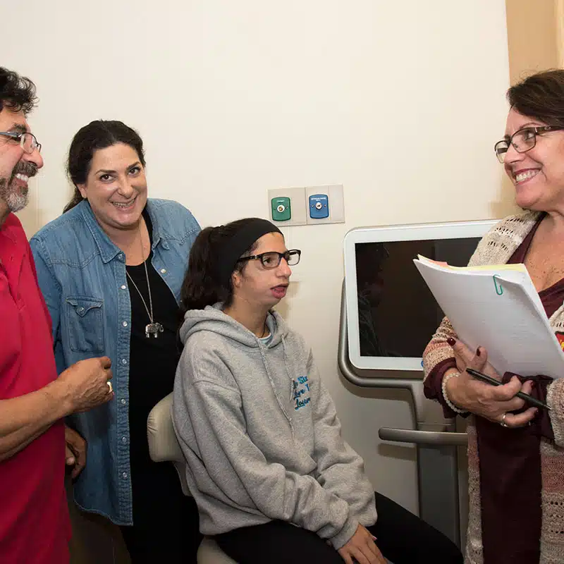
Helps us transform the lives of children and adults with facial differences.
Treatment Options
Managing Apert syndrome typically involves a multidisciplinary approach aimed at addressing the complex array of craniofacial, skeletal, and developmental abnormalities associated with the condition. Treatment modalities may include surgical interventions, supportive therapies, and ongoing medical management tailored to the specific needs of each individual.
Surgical Interventions
- Cranial Vault Remodeling: Surgical correction of craniosynostosis aims to release fused skull bones and reshape the skull to alleviate intracranial pressure and improve cranial aesthetics.
- Facial Reconstruction: Surgical procedures may be performed to address midfacial hypoplasia and other facial anomalies, such as advancement of the midface and reshaping of the orbits to improve facial symmetry and function. Fronto Orbital Advancement and Le Fort III surgery are common procedures for those born with Apert syndrome.
- Limb Reconstruction: Surgical correction of syndactyly and other limb abnormalities may involve separating fused digits, reconstructing finger and toe alignment, and optimizing hand and foot function.
Orthodontic and Dental Care
Orthodontic treatment may be necessary to address dental malocclusions and facilitate optimal dental and oral health. Regular dental examinations and preventive dental care are essential to monitor dental development and prevent complications such as dental crowding and malocclusion.
Psychosocial Support
Psychosocial support, including counseling and support groups, can play a crucial role in addressing the emotional and psychological needs of individuals with Apert syndrome and their families. Coping with the challenges associated with the condition and promoting self-esteem and social integration are important aspects of comprehensive care.
Learn more about myFace’s Support Group Options
Common questions about Apert Syndrome
How is Apert syndrome inherited?
Apert syndrome is typically inherited in an autosomal dominant pattern, meaning a child only needs to inherit one copy of the mutated gene from either parent to develop the condition.
Does Apert syndrome affect intelligence?
Apert syndrome primarily affects physical characteristics, such as the shape of the skull and limbs. While Apert syndrome itself does not directly impact intelligence, mental development can be affected by the skull’s impact on the brain. 60-70% of children with Apert syndrome have reduced IQ.
How common is Apert syndrome?
Apert syndrome is considered rare, affecting approximately 1 in every 65,000 to 88,000 births.
Patient Spotlight
Meet Sara. She’s found community through surfing and encourages other young people to find hobbies they enjoy.
“My lasting wish is that all children with craniofacial conditions or disabilities find the strength to fight for their dreams and live outside their comfort zone.”

For nearly 70 years, myFace has worked with patients and families to provide comprehensive care and support. In addition to providing access to state-of-the-art medical care, myFace provides other important services such as emotional support groups, workshops and educational webinars that offer guidance, counsel and resources for patients, families and the greater craniofacial community across the country.